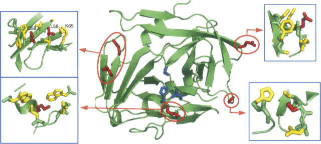
Figure 1.
Structure of the TEV protease. (Red) The residues subject to mutation (K45, L56, Q58, E106, S135). The four pictures correspond to the neighborhood of these five residues. (Blue) The catalytic triad H46, D81, and C151. The figure was generated with the PyMOL viewer (http://sourceforge.pymol.org).

