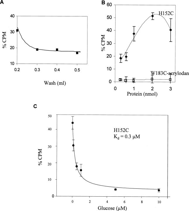
Figure 2.
Experiments were performed to establish screening parameters using purified proteins immobilized on Co2+ resin and challenged with a 10 μM glucose/3H-glucose solution. (A) By titrating the wash solution it was determined that 0.4 mL of DPBS removed 81% of the 3H-glucose from immobilized H152C. (B) The amount of purified protein needed to produce a large differential signal between the high-affinity H152C and W183C-acrylodan (Kd = 6 mM) was determined. The graph demonstrates that at 2 nmol of H152C there was a 13-fold signal increase over W183C-acrylodan. (C) Using the conditions above, immobilized H152C was challenged with 3H-glucose solution spiked with increasing amounts of cold glucose and determined a glucose affinity of 0.3 μM ± 0.05.

