
Figure 4.
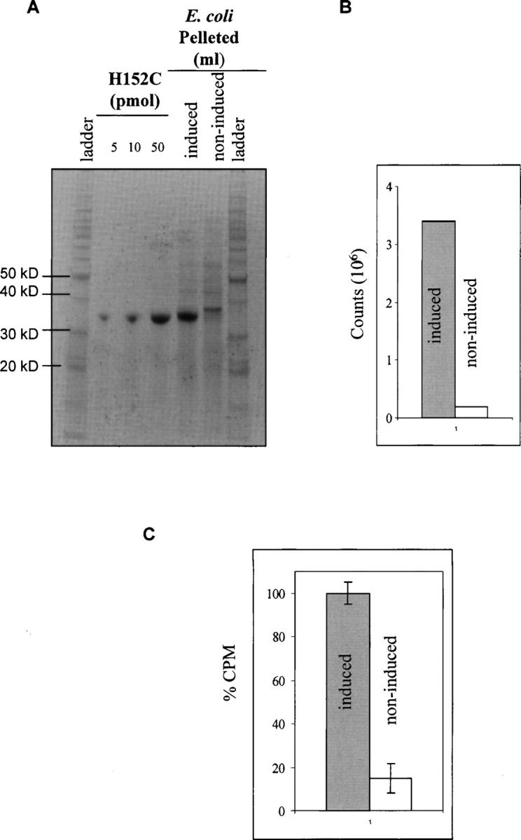
Testing GGBP binding of a 3H-glucose solution in bacteria. (A) Known amounts of purified H152C were analyzed by SDS-PAGE and compared to a 5 μL aliquot from 0.5 mL of bacteria that had been lysed into 100 μL. The bacteria from both induced and noninduced samples were run side-by-side. (B) With background correction the noninduced sample signal was ∼24-fold less than the induced. (C) Induced and noninduced E. coli samples were run through the screening protocol. The induced sample demonstrated 5.6-fold greater signal than the noninduced.