
Fig. 2.
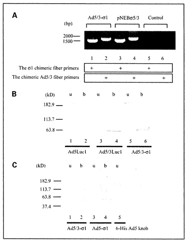
Analysis of fibers in rescued vps. A, detection of fiber genes in the adenovirus genome. Rescued vps were analyzed with PCR using pairs of the σ1 chimeric fiber primers or the chimeric Ad5/3 fiber primers. PNEBσ5/3 was used as a positive control for both fibers. Absence of a PCR template was designated as the “Control.” B and C, Western blot analysis of fiber proteins in purified virions. B, a total of 1.0 × 10¹⁰ vp per lane of Ad5Luc1 with the wild-type Ad5 fiber (lanes 1 and 2), Ad5/3Luc1 with the chimeric Ad5/3 fiber (lanes3 and 4), or Ad5/3-σ1 with dual fibers (lanes 5 and 6) was resuspended in Laemmli buffer before SDS-PAGE, electrotransferred, and detected with the 4D2 anti-Ad5 fiber tail antibody. The samples in lanes 2,4, and 6 were boiled (b), whereas lanes 1, 3, and 5 [unboiled (u)] contain proteins in their native trimeric configuration. C, a total of 1.0 × 10¹¹ vp per lane of Ad5/3-σ1 (lanes 1 and 2) and Ad5-σ1 (lanes 3 and 4) with dual fibers was probed with an anti-6-His antibody. Lane 1, unboiled Ad5/3-σ1 virions; lane 2, boiled Ad5/3-σ1 virions; lane 3, unboiled Ad5-σ1 virions; lane 4, boiled Ad5-σ1 virions; lane 5, recombinant Ad5 knob with a 6-His tag as a positive antibody control. We consider the protein band appearing at~113 kDa to be the dimeric form of the σ1 chimeric fiber.