
Figure 3.
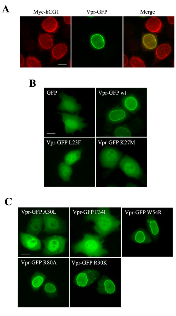
Subcellular distribution of the Vpr mutants. A) Colocalization of Vpr and hCG1 at the NE. HeLa cells co-expressing Vpr-GFP (middle row) and Myc-hCG1 (left row) fusion proteins were permeabilized with digitonin, fixed, and subsequently stained with an anti-Myc monoclonal antibody. B and C) Localization of wt and mutated Vpr-GFP fusions. HeLa cells expressing either GFP, wt Vpr-GFP, or the indicated Vpr-GFP mutants were fixed and directly examined. Cells were analyzed by epifluorescence microscopy, and images were acquired using a CCD camera. Scale bar, 10 μm.