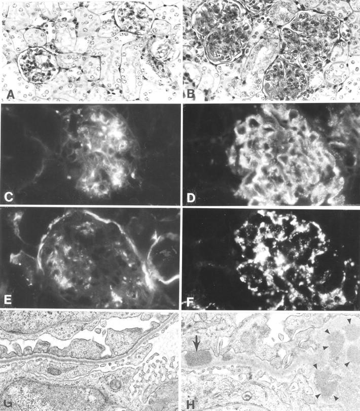
Figure 3.

Histological, immunofluorescence, and EM studies of representative kidneys from NZM.C57Lc1 and NZM.C57Lc4 female mice. (A) Normal glomeruli (hematoxylin and eosin staining, ×200) are seen in NZM.C57Lc1. (B) In contrast, in the NZM.C57Lc4 congenic, enlarged glomeruli with mesangial proliferation, hypercellularity, obliterated capillary loops, and glomerulosclerosis are evident. (C) Immunofluorescence studies show some mesangial IgG deposits in NZM.C57Lc1, similar to the pattern seen in aged C57L/J. (D) A coarsely granular staining pattern of IgG deposits in both the mesangia and peripheral capillary walls of the glomeruli of NZM.C57Lc4. (E) Staining of the Bowman capsule and mesangia with anti-C3 Ab are seen in NZM.C57Lc1. (F) Coarsely granular staining by anti-C3 Ab throughout the glomeruli is seen in NZM.C57Lc4. (G) EM study shows normal glomeruli without electron-dense deposits in the subepithelial or subendothelial spaces (×10,000) in the kidney of NZM.C57Lc1. (H) In comparison, electron-dense deposits in both subendothelial space (arrow) and the mesangia (arrowheads) in the glomeruli of NZM.C57Lc4 are readily detected.
