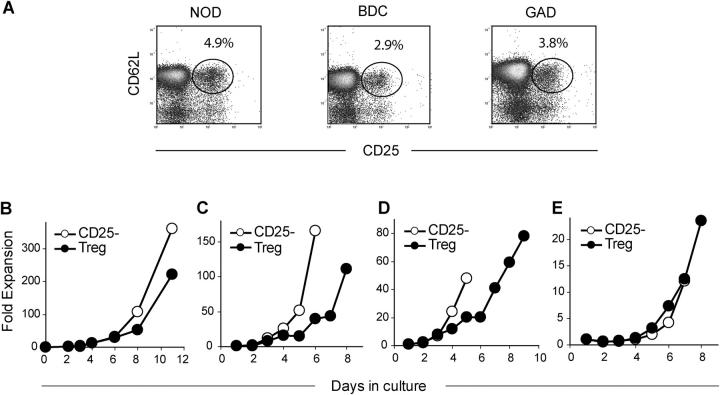
Figure 1.
In vitro expansion of Tregs. (A) Representative flow cytometry plots of CD25 and CD62L expression on CD4 cells from NOD (left), BDC2.5 (middle), and GAD286 (right) mice. FACS®-purified Tregs (•) and CD4+ CD62L+ CD25− cells (○) from NOD (B), BDC2.5 TCR Tg (C), or GAD286 TCR Tg (D) mice were stimulated in vitro with anti-CD3– and anti-CD28–coated beads along with IL-2. (E) T cells from BDC2.5 TCR Tg mice were expanded as described above with p31-linked IAg7-mIgG2a immobilized on latex beads. All cultures were quantitated by viable cell counting.

