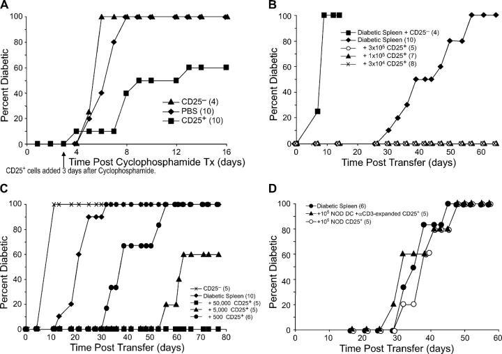
Figure 5.
Expanded CD25+ CD4+ T cells function in vivo to suppress development of diabetes. (A) 4–6-wk-old NOD.BDC2.5 mice were given cyclophosphamide i.p. 3 d later, either 5 × 10⁵ DC-expanded CD25+ CD4+ T cells or CD25− CD4+ cells were injected i.v. (B) NOD.scid females were injected with 3 × 10⁶ spleen cells from a diabetic NOD female and either nothing or the indicated numbers of DC-expanded CD25+ CD4+ T cells or 3 × 10⁵ CD25− CD4+ cells from BDC2.5 mice. (C) NOD.scid females were injected with either 4 × 10⁵ CD25− CD4+ cells from BDC2.5 mice, or 8 × 10⁶ spleen cells from a diabetic NOD female and either nothing or the indicated numbers of DC-expanded CD25+ CD4+ T cells from BDC2.5 mice. The difference between diabetic spleen alone to diabetic spleen plus 500 CD25+ CD4+ cells was significant (P = 0.002), as was diabetic spleen to diabetic spleen plus 5,000 CD25+ CD4+ cells (P = 0.002). One representative result from two experiments is shown. (D) NOD.scid females were injected with 8 × 10⁶ diabetic spleen cells alone or with 105 freshly isolated or DC/αCD3-expanded CD25+ CD4+ T cells from NOD mice. The number of mice in each group is indicated in parentheses.

