
Figure 2.
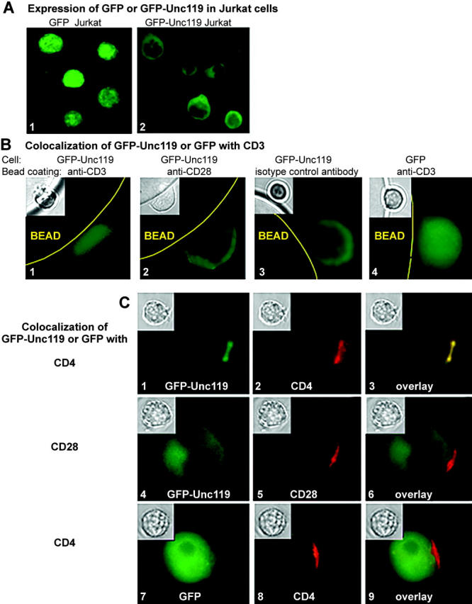
Unc119 translocates to the TCR complex upon anti-CD3 stimulation. (A) Expression of GFP-Unc119 fusion protein. Jurkat cells were stably transfected with pLEGFP-N1 containing cDNA for GFP alone (1) or GFP-Unc119 fusion protein (2) and examined by fluorescence microscopy. (B) Unc119 translocates to CD3 upon stimulation. GFP-Unc119 (1, 2, and 3) or GFP (4) Jurkat cells were incubated with anti-CD3 (1 and 4) or anti-CD28 (2) or isotype-control antibody (3)–coated protein A/G agarose beads, fixed, and analyzed by fluorescence and light (inset) microscopy. (C) Unc119 translocates to CD4 upon stimulation. Receptors of GFP-IRIP (1–6) or GFP (7–9) Jurkat cells were capped with anti-CD4 (1–3, 7–9), anti-CD28 (4–6), or isotype controls (not depicted) and examined by fluorescence or light (inset) microscopy. (left) GFP/GFP-IRIP fluorescence. (middle) Rhodamine (CD4 or CD28) fluorescence. (right) Color overlay.