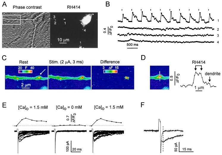
Figure 2.
Bouton activation by extracellular electrical stimulation is highly spatially selective. (A) Phase-contrast and RH414 image of an area comprising several boutons. (B) A train of short, depolarizing pulses (1.5 μA, 2 ms, 0.2 Hz) evoked a Ca2+ elevation only in the stimulated terminal (arrow). All other RH414-stained boutons in the view field lacked stimulus-locked Ca2+ transients (not all of them shown). Binning, 2 × 2; exposure time, 50 ms. (C) The same experiment. Enlarged image of the boxed area in A. The differential display illustrates selective activation of the stimulated bouton. (D) Profile of fluorescence change along a line through the stimulated bouton. Arrows point to the margins of the RH414-stained zone and the underlying dendrite. The stimulus (2 μA, 2 ms) increased the relative fluorescence by 50–60% in the bouton and by 5–10% in the dendrite. (E) Experiment with local application of Ca2+-free solution. Stimulus intensity was 1.5 μA in standard extracellular [Ca2+] and was varied from 1.5 to 4 μA in Ca2+-free solution. (Upper) Averaged [Ca2+]pre. (Lower) Superposition of postsynaptic responses. Note rapid and almost intensity-independent poststimulus relaxation in failure traces during the Ca2+-free test. (F) Single, enlarged failure trace and eIPSC in response to identical stimuli (1.5 μA, 2 ms).

