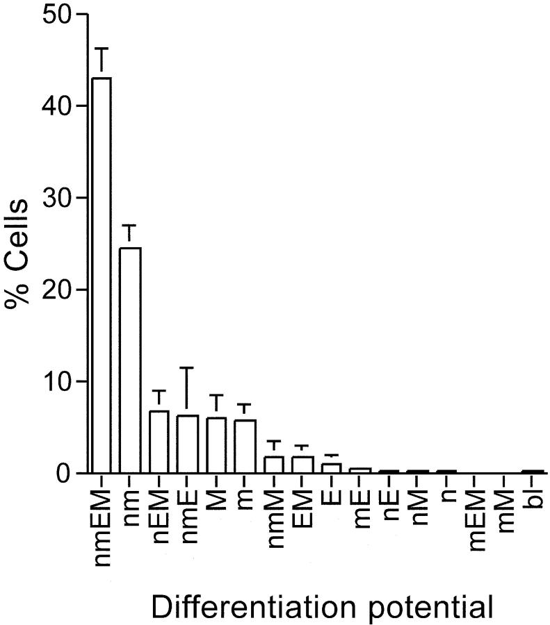
Figure 2.
Colony-forming ability of single CD34−KSL cells. CD34− KSL cells were individually cultured in the presence of SCF, IL-3, TPO, and EPO for 2 wk. Percentages of CFCs with different differentiation potentials are shown based on three independent experiments. Colony cells were morphologically identified as neutrophils (n), macrophages (m), erythroblasts (E), or megakaryocytes (M). Otherwise, unidentified immature cells were designated as blastlike cells (bl). The nmEM cells constituted 43.2 ± 3.2% (mean ± SD; n = 3) of the colony-forming CD34−KSL cells.

