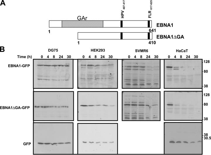
Figure 1.
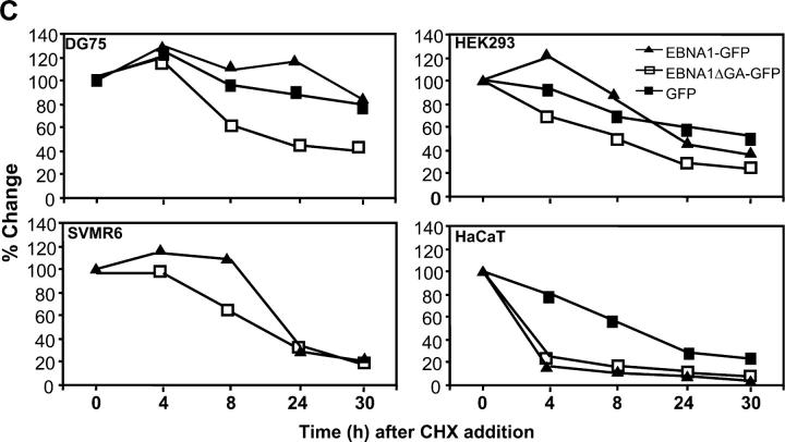
(A) Schematic description of EBNA1 and EBNA1ΔGA expression constructs showing localization of FLR and HPV epitopes. (B) Intracellular degradation of EBNA1-GFP in different cell types. DG75 B cells, HEK293 epithelial cells, SVMR6 keratinocytes, and HaCaT keratinocytes were transfected with expression constructs EBNA1-GFP, EBNA1ΔGA-GFP, or the control plasmid pEGFP-N1. At 36 h after transfection, the cells were degraded over a 30-h time course in the presence of 50 μg/ml cycloheximide as described in Materials and Methods. Molecular weight standards are indicated at the side of each panel. (C) Densitometric analysis of EBNA1-GFP, EBNA1ΔGA-GFP, and GFP expression. Band intensities were quantified by analysis of the imaging data and plotted as a relative percentage of the signal at time 0 for EBNA1-GFP, EBNA1ΔGA-GFP, and GFP.


