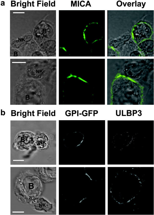
Figure 3.

Recruitment of ULBP3 and MICA to the NK cell immune synapse. (a) CD56+ CD3− human peripheral blood NK cells were coincubated with Daudi/Class I+/MICA cells for 15 min, fixed, and stained with anti-MICA mAb, and the distribution at the immunological synapse was determined. The first row shows a single NK cell forming two activating synapses with two different Daudi/Class I+/MICA cells. Data shown are representative of three experiments in which 301 NK-Daudi/Class I+/MICA conjugates were assessed. (b) A CD56+ CD3− human peripheral blood NK clone was coincubated with GPI-GFP–expressing Namalwa transfectants for 15 min, fixed, and stained for ULBP3. Images are representative of 13 out of 14 conjugates where ULBP3 was clearly seen to accumulate at the synapse. Bars, 5 μm.
