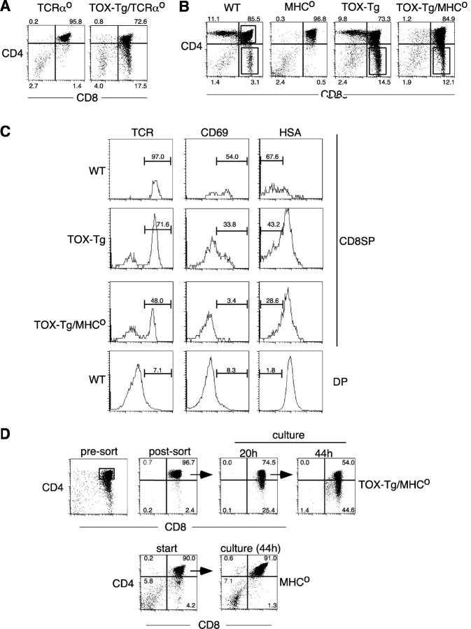
Figure 2.
TOX induces development of CD8SP thymocytes in the absence of TCR–MHC interactions. (A and B) Expression of CD4 and CD8α on thymocytes from mice with the indicated genotype. Percentages of total thymocytes in each quadrant are indicated. (C) Expression of TCRβ, CD69, and HSA on CD8SP thymocytes gated as in B from mice with the indicated genotypes. DP thymocytes from wild-type mice were used as a control to set appropriate gates. The percentage of TCRhi, CD69+, and HSAlo cells are shown. (D) Total MHCo thymocytes or sorted DP thymocytes from TOX-Tg/MHCo mice were cultured ex vivo and analyzed for CD4 and CD8α expression by flow cytometry after 20 or 44 h. Percentages of total thymocytes in each quadrant are shown. Viable yield of TOX-Tg/MHCo thymocytes was 73% after 20 h of culture.

