Figure 4.
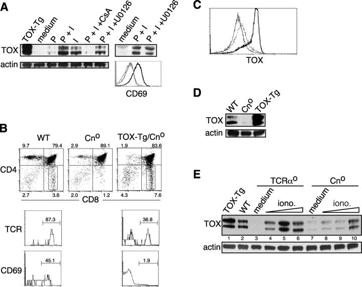
Expression of TOX is regulated by Cn activation. (A) Expression of TOX in TCRαo thymocytes activated with PMA and ionomycin in the absence or presence of inhibitors of Cn (cyclosporin A, CsA) or MEK (U0126). Also shown is CD69 expression on cells cultured in medium (thin line), with PMA and ionomycin (thick black line), or with PMA/ionomycin and U0126 (thick gray line). Expression of TOX in thymocytes derived from a TOX-Tg mouse is shown as a control. (B) Expression of CD4 and CD8α on thymocytes derived from various animals as indicated (dot plots). Percentage of total thymocytes in each quadrant is shown. Expression of TCRβ and CD69 was also analyzed on gated CD8SP thymocytes, and the percentage of TCRhi or CD69+ cells is indicated. Expression of TOX was determined by intracellular staining and flow cytometry (C) in WT (dashed line), Cno (thin line), and TOX-Tg (thick line) thymocytes or by Western blot of whole cell lysates (D). (E) Thymocytes from TCRαo and Cno mice were cultured in medium (lanes 3 and 7) or 0.2 ng/ml of PMA and 0.05 (lanes 4 and 8), 0.1 (lanes 5 and 9), or 0.2 (lanes 6 and 10) μg/ml of ionomycin. TOX-Tg thymocytes were analyzed as a control (lane 1).

