
Figure 5.
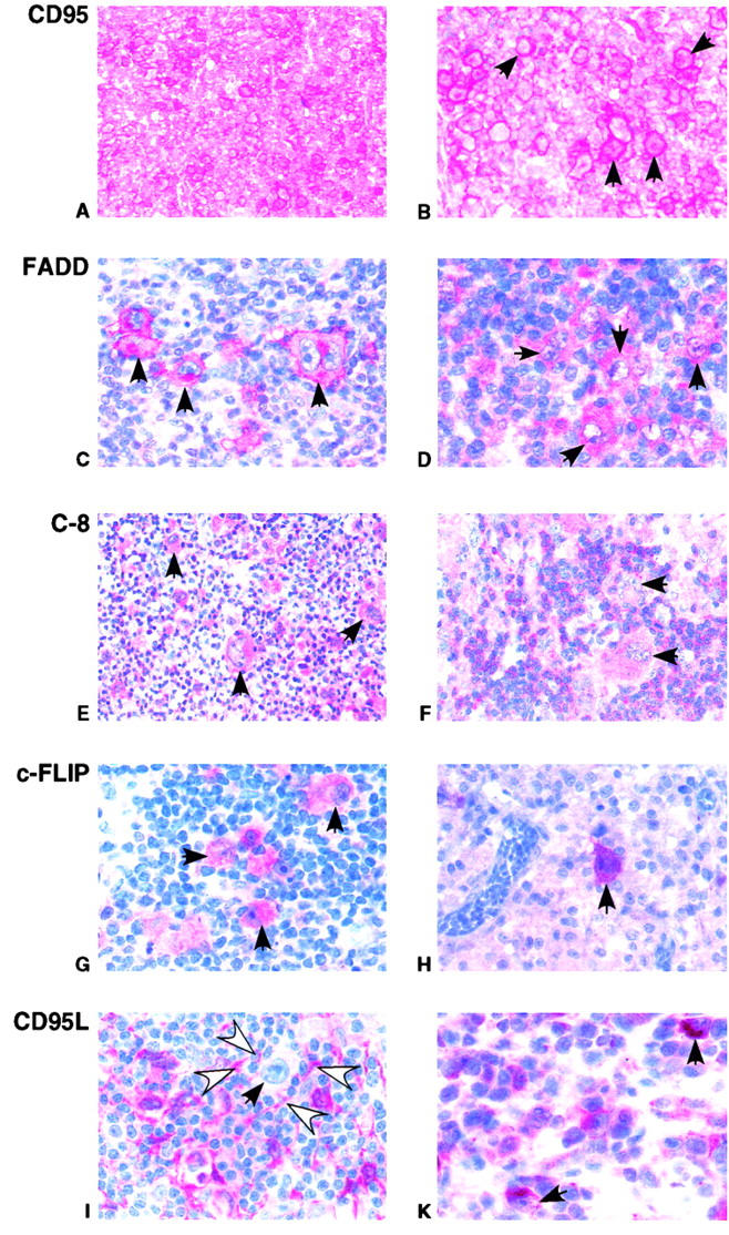
Expression patterns of CD95, FADD, C-8, c-FLIP, and CD95L in HRS cells of cHL. Immunohistochemistry of representative biopsy specimen. (A and B) Strong CD95 expression in HRS cells (exemplarily marked by arrows). (C) Strong FADD expression in HRS (arrows), but not in surrounding cells. (D) Moderate FADD expression in HRS cells (arrows). (E and F) C-8 expression of two representative cases. (E) HRS cells (arrows) stain at least as strong as surrounding cells for C-8. (F) HRS cells stain in a less intense fashion than surrounding cells. (G and H) Strong c-FLIP expression in HRS cells (arrows), but not surrounding cells. (I and K) CD95L expression in cHL. (I) In the majority of cases, HRS cells (solid arrow) do not stain for CD95L. In contrast, surrounding cells (open arrows) stain for CD95L. (K) In one case, strong expression of CD95L was detectable in HRS cells (arrows).