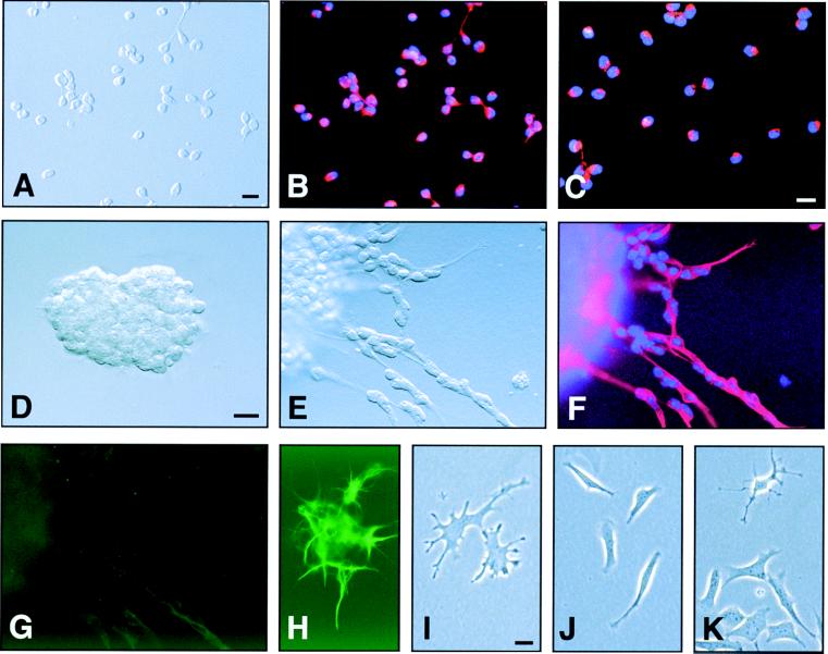
Figure 2.
Fractionation of SVZ cells by differential adhesion yields populations of cells with distinct characteristics. (A–C) Fraction 1, type A cells. See Results and Fig. 3 for details. (A) DIC image of isolated type A cells. (B) Epifluorescent image showing Tuj1 staining (red). (C) Purified type A cells are PSA-NCAM+ (red) and can migrate in chains. (D) Aggregate of type A cells immediately after they were embedded in Matrigel. (E) Chain migration from aggregates after 6 h in culture. The culture in E was double-stained for Tuj1 (F) and GFAP (G). (H) A positive control for GFAP staining (green) in Matrigel culture. (I–K) Fraction 4, B/C cells. Most of the adherent cells had a flattened, spread, phase-dark appearance, and ≈30% of these cells were GFAP+ (see Fig. 3 Inset). Nuclei are counterstained with Hoechst 33258 (blue). (Bar = 10 μm.)

