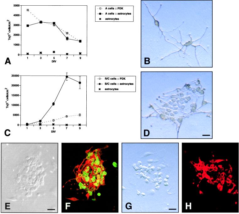
Figure 4.
Fraction 1 (type A cells) and fraction 4 (type B/C cells) in astrocyte coculture. (A) Time course of a cell–astrocyte coculture vs. culture on PDK. Error bars = SEM of triplicate cultures. (B) Tuj1+ cells stained with diaminobenzidine in a cell–astrocyte coculture at 5 DIV. (C) Time course of B/C cell–astrocyte coculture vs. culture on PDK. (D) Typical colony of Tuj1+ cells stained with diaminobenzidine in astrocyte coculture at 5 DIV. (E and F) Cocultures were exposed to BrdUrd from 2 to 4 DIV. (E) DIC image of a neuronal colony. (F) Same colony double-stained for Tuj1 (red) and BrdUrd (green). DIC (G) and epifluorescent (H) images of a neuronal colony stained for PSA-NCAM. (Bars = 10 μm.)

