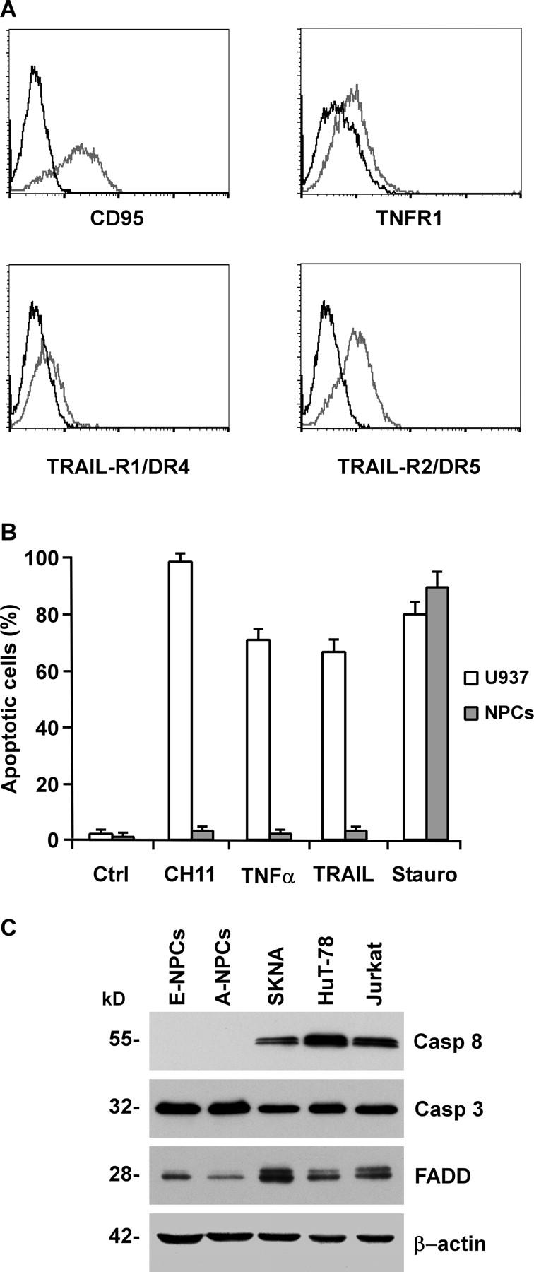
Figure 1.

Expression of DRs and sensitivity to apoptosis in tissue-isolated embryonic and adult NPCs. (A) Flow cytometry analysis of DRs in NPCs. Cells were stained with antibodies specific to CD95, TNFR1, TRAIL-R1/DR4 and TRAIL-R2/DR5 (gray), or control IgGs (black). (B) Percentage of apoptotic cells in NPCs untreated (Ctrl) or exposed for 24 h to 500 ng/ml anti-CD95 (CH11), 500 U/ml TNF-α, 500 ng/ml TRAIL, or 0.25 μM staurosporine (Stauro). The human monocytic cells U937 were used as a positive control. The results are the mean ± SD of four independent experiments. (C) Immunoblot analysis of caspase 8 (Casp 8), caspase 3 (Casp 3), and FADD on embryonic (E-NPCs) and adult NPCs (A-NPCs) as compared with control cell lines. Detection of β-actin in the same membrane blot served as loading control. One representative experiment out of four is shown.
