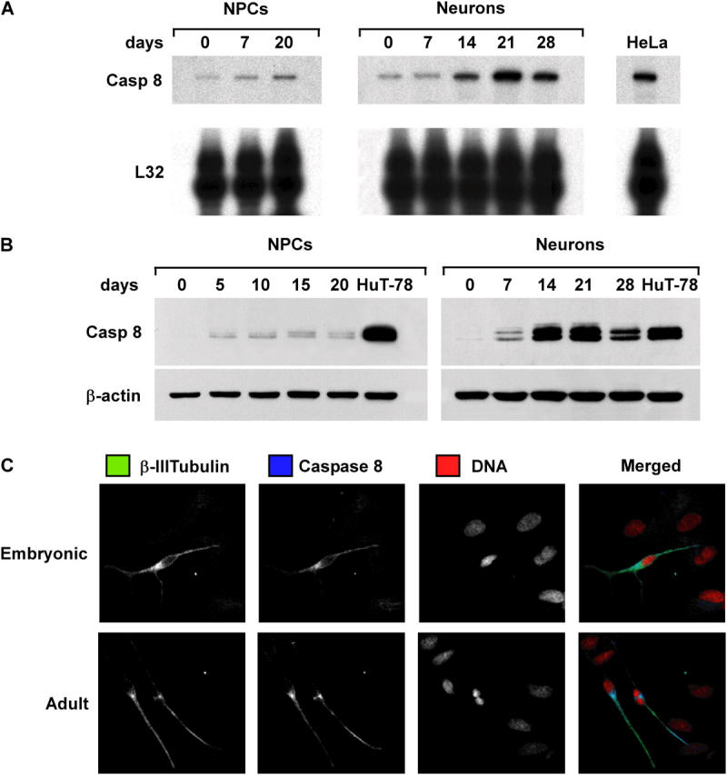
Figure 2.

Expression of caspase 8 during NPC and NT2/D1 differentiation. (A) RNase protection assay for caspase 8 mRNA in NPCs and NT2-neurons at different days of differentiation. L32 levels were used as loading control, and RNA of HeLa cells were used as reference control. (B) Immunoblot analysis of caspase 8 expression in cells treated as in A. Cell lysates of HuT-78 cells were used as control. (C) Cells from differentiated embryonic and adult NPCs were analyzed by confocal microscopy to detect β-III tubulin (green), caspase 8 (blue), and DNA (red). One representative experiment out of four performed with commercial and tissue-isolated NPCs is shown.
