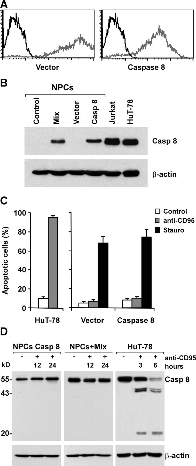
Figure 4.

Absence of sensitization to CD95-induced apoptosis by exogenous caspase 8 expression in adult NPCs. (A) Flow cytometry profiles of GFP-positive NPCs transduced with empty Tween (Vector) or Tween/Caspase 8 (Caspase 8) vector (gray) as compared with nontransduced NPCs (black). (B) Immunoblot analysis of caspase 8 and β-actin in NPCs transduced as in A or treated with TNF-α, IFN-γ, and IL-1β (Mix). Jurkat and HuT-78 cell lines were used as positive controls. (C) Percentage of apoptosis in NPCs transduced as in A and treated with anti-CD95 or staurosporine. HuT-78 cells were used as positive control. The results are the mean ± SD of four independent experiments. (D) Immunoblot analysis of caspase 8 and β-actin in NPCs cultured with or without inflammatory cytokines and exposed to anti-CD95 for 12 or 24 h. In the control cell line HuT-78, the proteolytic products of caspase 8 were detectable shortly after CD95 stimulation.
