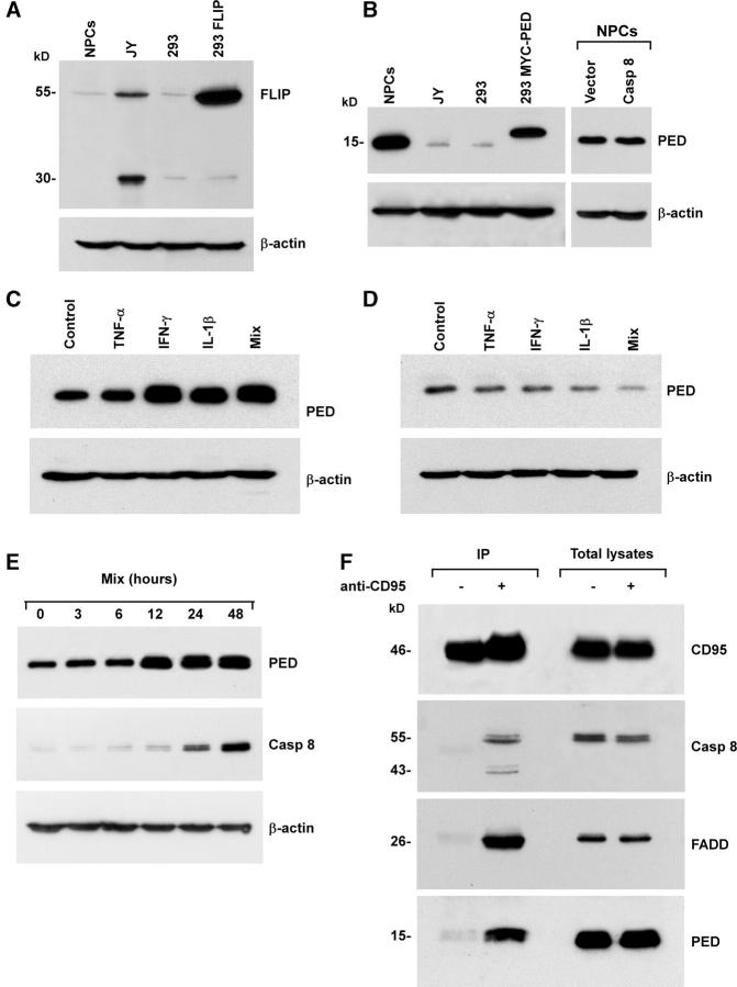
Figure 5.
Expression of DISC inhibitory proteins in tissue-isolated embryonic and adult NPCs. Immunoblot analysis of cFLIP (A) and PED/PEA-15 (B) on NPCs, JY, and 293T cell lines, the latter transfected with Tween/cFLIP (293 FLIP) or Tween/MYC-PED/PEA-15 (293 MYC-PED), were used as positive control. Immunoblot analysis of PED/PEA-15 in NPCs (C) and NT2-neurons (D) untreated (Control) or treated with TNF-α, IFN-γ, and IL-1β alone or in combination (Mix). One representative experiment out of three is shown. (E) Immunoblot analysis of PED/PEA-15 and caspase 8 in NPCs at different time points after stimulation with inflammatory cytokines. Detection of β-actin in the same membrane blot served as loading control. (F) Immunoprecipitation (IP) of the CD95 DISC in control (−) or CD95-stimulated (+) NPCs treated with TNF-α, IFN-γ, and IL-1β. CD95 was immunoprecipitated and blot probed for CD95, caspase 8, FADD, and PED/PEA-15. One representative experiment out of four is shown.

