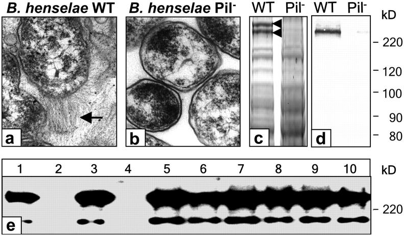
Figure 1.
Phase variation of B. henselae. (a and b) Expression of so-called type IV-like pili (BadA, arrow; see also Fig. S1, available at http://www.jem.org/cgi/content/full/jem.20040500/DC1) of B. henselae Marseille WT not detectable on the surface of B. henselae Pil−. (c) SDS-PAGE of B. henselae WT and Pil− showing two differential HMW bands. The lower band is Fn (240 kD), the upper is BadA (calculated mass: 340 kD). (d) Detection of Fn binding of B. henselae WT and Pil− by Western blotting. Bacteria-bound Fn was detected using an anti-Fn antibody. (e) Screening of a B. henselae transposon library for Fn binding. B. henselae WT (lane 1), Pil− (lane 2), and transposon mutants (lanes 3–10). Fn binding is not detectable in B. henselae Pil− and in a transposon mutant (lane 4), suggesting loss of BadA (pilus) expression.

