Figure 4.

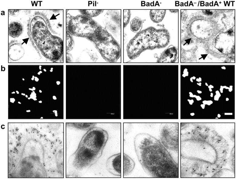
Complementation of B. henselae BadA−. (a) Detection of BadA expression in B. henselae WT, Pil−, BadA−, and BadA−/BadA+ WT by TEM. Note the brush-like arrangement of BadA on the surface of the WT and the complemented mutant (arrows) missing in the Pil− variant and the BadA− mutant. (b) Detection of BadA expression by immunofluorescence using an anti-BadA rabbit serum. Bar, 2 μm. (c) Detection of BadA expression by IEM using 10 nm gold-conjugated goat anti–rabbit IgG.
