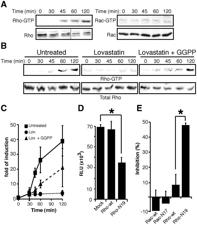
Figure 3.
Statins inhibit HIV-1 infection by down-regulating Rho activation. (A) Serum-starved MT2-CCR5 cells were incubated with HIV-1 and cell lysates were assayed for active Rho or Rac. Total Rho or Rac was analyzed in parallel in crude cell extracts as a protein loading control. One experiment out of three is shown. Black line indicates that different sections of the same gel were juxtaposed. (B) Active Rho was determined in untreated, Lov-, or Lov plus GGPP–treated cells, as described above. Total Rho in crude cell extracts is shown as a loading control. One representative experiment out of three is shown. (C) Western blots from three independent experiments as in B were quantified by densitometry and values were normalized using Rho in crude cell extracts as a loading control. Data points are plotted relative to mean values of cells not exposed to virus (time 0) for each condition. (D) Single-round infections of MT2-CCR5 cells transfected with mock, wild-type Rho, or mutant Rho-N19 using an HIV-1Ada–pseudotyped, replication-defective virus. *, P < 0.05. Kruskal-Wallis test. (E) HeLa-CD4 cells transfected with wild-type Rac, wild-type Rho, Rac-N17, or Rho-N19 were mixed with HIV gp160–expressing BSC40 cells. Cell fusion events were measured and normalized relative to mock transfected cells. *, P < 0.05. Kruskal-Wallis test. (D and E) Data are mean ± SD of duplicate points (n = 3).

