
Figure 1.
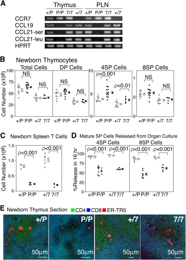
Characterization of thymuses of newborn CCR7- or CCR7L-deficient mice. (A) RT-PCR analysis of lymphoid organs in CCR7- or CCR7L-deficient mice. Total cellular cDNA of thymuses and peripheral lymph nodes (PLN) was isolated from +/plt (+/P), plt/plt (P/P), CCR7+/− (+/7), and CCR7−/− (7/7) mice and was PCR amplified for CCR7, CCL19, CCL21-ser, CCL21-leu, and HPRT. (B) Thymocytes from indicated newborn mice at 5 d old were stained for CD4, CD8, and TCRβ and analyzed by flow cytometry. DP, 4SP, and 8SP represent CD4+CD8+, CD4+CD8−TCRβhigh, and CD4−CD8+TCRβhigh, respectively. Data from individual mice and means ± SE are indicated. (C) CD3highTCRβhigh T cells in the spleen were measured in 5-d-old mice. (D) Newborn mouse thymus lobes were cultured for 16 h, and cells within and outside the thymus lobes were stained for CD4 and CD8. Cells that were released from the thymus lobes were measured for CD4+CD8− and CD4−CD8+ populations. (E) Three-color immunofluorescence analysis of newborn mouse thymus sections on the day of birth for CD4, CD8, and ER-TR5. Cells in cyan color (merging of green and blue) indicate CD4+CD8+ cells expressing both CD4 (in green) and CD8 (in blue). p values were calculated by the Student's t test. NS, not significant.