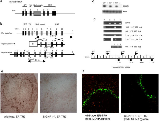
Figure 1.
Inactivation of the mouse SIGN-R1 gene by homologous recombination. (a) Structure of the human DC-SIGN locus. (b) Structure of the mouse SIGN-R1 locus, the targeting vector, and the predicted homologous recombination event are shown. NeoR, neomycin resistance cassette; TK, thymidine kinase cassette; B, BamHI. (c) RT-PCR analysis of SIGN-R1 expression. RNA was prepared from splenocytes and PCR was performed. −/−, SIGN-R1−/−. (d) RT-PCR analysis of SIGN-R1 expression using PCR primers throughout the cDNA to verify gene deletion. (1) Wild-type. (2) SIGN-R1−/−. (3) Wild-type. (4) SIGN-R1−/−. (e) Staining with ER-TR9 (anti–SIGN-R1) monoclonal antibody using horseradish peroxidase detection. (f) Co-staining with ER-TR9 monoclonal antibody (Texas red) and MOMA monoclonal antibody (FITC) (identifying marginal metallophilic macrophages). Original magnification, 10.

