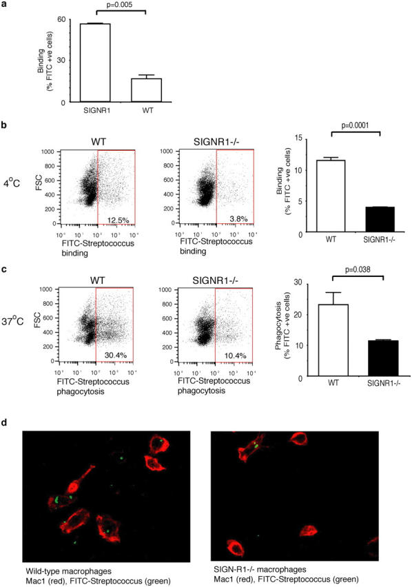
Figure 5.

Impaired recognition of S. pneumoniae by SIGN-R1−/− peritoneal macrophages. (a) S. pneumoniae bind preferentially to SIGN-R1–transduced NIH3T3 (SIGNR1) cells compared with wild-type (WT) NIH3T3. Cells were incubated for 1 h at 4°C with FITC-labeled pneumococci, washed, and analyzed by flow cytometry. Values represent the mean of triplicates, and p-values were obtained using an unpaired Student's t test. (b–d) Pooled peritoneal macrophages from wild-type (n = 2) or SIGN-R1−/− mice (n = 2) were incubated for 30 min with FITC-labeled S. pneumoniae at 4°C to assess binding (b), or 37°C to assess phagocytosis (c), followed by flow cytometric analysis. Binding or phagocytosis are expressed as percentage of FITC positive cells. SIGN-R1−/− peritoneal macrophages show reduced binding and phagocytosis of S. pneumoniae. Representative FACS profiles are shown. Values represent mean of triplicates, the experiments shown are representative of two, and p-values were obtained using an unpaired Student's t test. (d) Confocal images of wild-type and SIGN-R1−/− peritoneal macrophages labeled with Mac-1 allophycocyanin after incubation with FITC-labeled S. pneumoniae at 37°C for 45 min.
