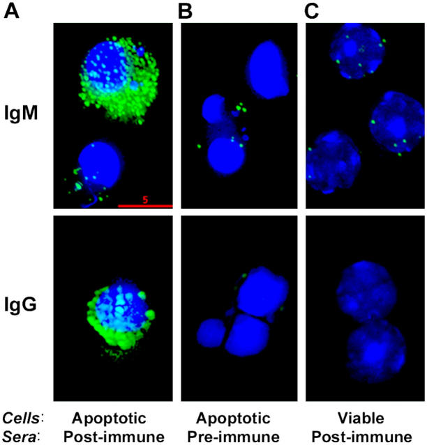
Figure 4.
Immunofluorescence deconvolution microscopy of sera binding to apoptotic cells: Pooled (IgM) or a representative (IgG) preimmune and postimmune sera from NIH/Swiss-Webster mice immunized with apoptotic thymocytes were diluted 1:200 in PBS with 1% BSA and incubated with apoptotic or normal thymocytes. IgM or IgG binding to the cells was detected by fluorescein-conjugated F(ab)2 fragments against mouse IgM or IgG, respectively. (A) Note the marked binding of postimmune sera (green) to apoptotic thymocytes with their characteristic condensed, fragmented nuclei detected by Hoechst staining (blue). (B) Note the negative staining of preimmune sera to apoptotic cells and (C) postimmune sera to normal thymocytes. Bar, 5 μm.

