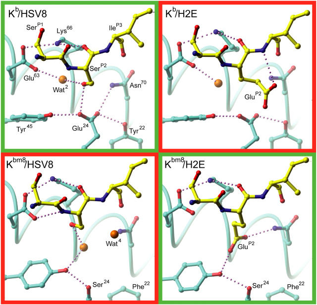
Figure 5.
pMHC B pocket hydrogen bonding schemes. The B pocket hydrogen bonding networks of all four pMHCs are depicted. The specific interactions of peptide P2 residues with MHC are highlighted. Recognized pMHCs are in green boxes and the unrecognized pMHCs are in red boxes. (cyan) MHC carbon atoms; (yellow) peptide carbon; (red) oxygen; (blue) nitrogen; (orange) water. Potential hydrogen bonds are depicted as small purple spheres.

