
Figure 2.
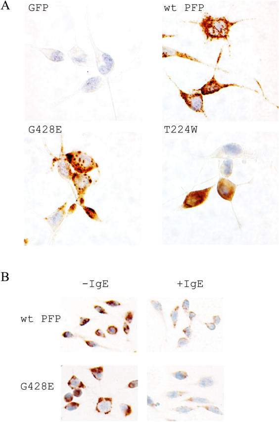
T224W and G428E perforin localize differently in RBL cells. (A) Immunohistochemistry of perforin-expressing RBL cells demonstrated with antiperforin antibody PI-8 and counterstained with eosin. (B) RBL cells either unlabeled or labeled with α-TNP–IgE were stained as in A, after degranulation was induced by transient incubation with TNP-labeled target cells. Magnification, 400×.