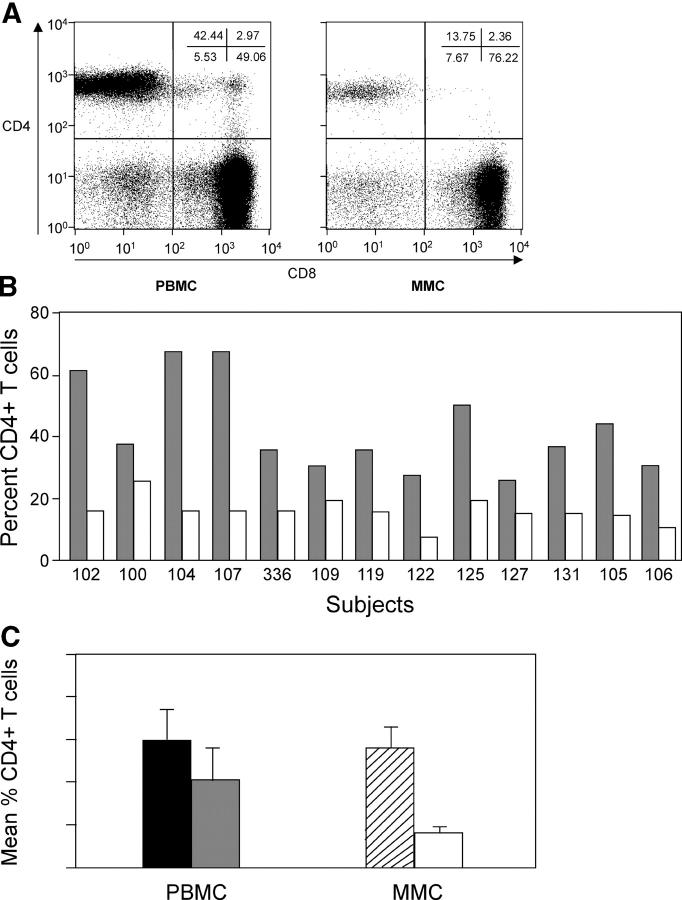
Figure 1.
CD4+ T cells are preferentially depleted in the GI tract in acute and early HIV-1 infection. PBMCs and MMCs from the rectosigmoid region from group 1, acute and early HIV-1 infection subjects (n = 13), and HIV-1–uninfected controls (n = 10) were analyzed by flow cytometry. CD3+ gated lymphocytes were analyzed for the expression of CD4 and CD8. (A) A representative flow plot from subject 105 is depicted. CD8+ T cells are shown on the x axis and CD4+ T cells are shown on the y axis. (B) Comparison of CD4+ T cells in the blood and GI tract of all 13 group 1 primary infection subjects. The percent of CD4+ T cells is shown in PBMCs (gray) and MMCs (white) per study subject. (C) Comparison of the mean percent of CD4+ T cells in the PBMCs of HIV-1–uninfected controls (black bar) and group1 subjects (gray bar), and MMCs of HIV-1–uninfected controls (hatched bar) and group 1 subjects (white bar).

