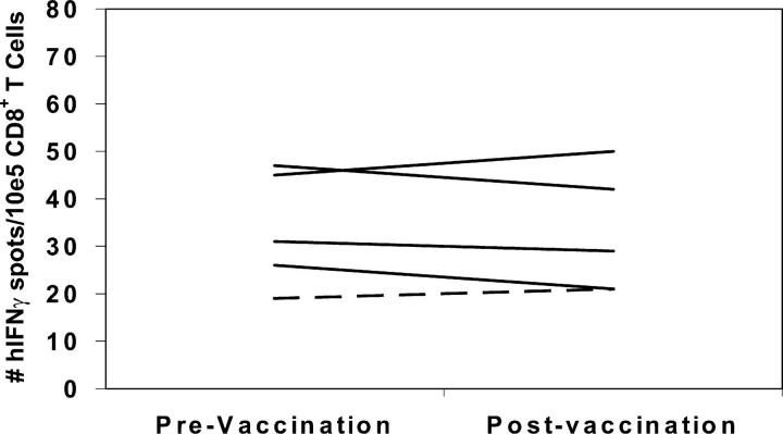
Figure 3.
ELISPOT analysis of CD8+ T cells from PBMCs demonstrates similar pre- and postvaccination responses to the influenza matrix protein HLA-A2 binding epitope M1 (GILGFVFTL) in all HLA-A2+ patients This analysis was performed on the same PBL samples described in Fig. 2. The DTH responders are represented by dotted lines, and the DTH nonresponders are represented by solid lines. For the detection of nonspecific background, the number of IFN-γ spots for CD8+ T cells specific for the irrelevant control peptides were counted. The HLA-A2 binding HIV-gag protein-derived epitope (SLYNTVATL), the HLA-A3 binding HIV-NEF protein-derived epitope (QVPLRPMTYK), and the HLA-A24 binding melanoma tyrosinase protein-derived epitope (AFLPWHRLF) were used as negative control peptides in these assays. Data represent the mean of each condition assayed in triplicate, and standard deviations were <5%. The number of human IFN-γ spots per 105 CD8+ T cells is plotted. Analysis of each patient's PBLs was performed at least twice, and all ELISPOT assays were performed in a blinded fashion.

