Figure 4.
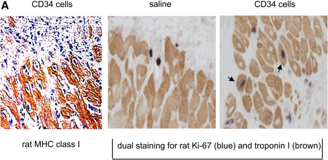
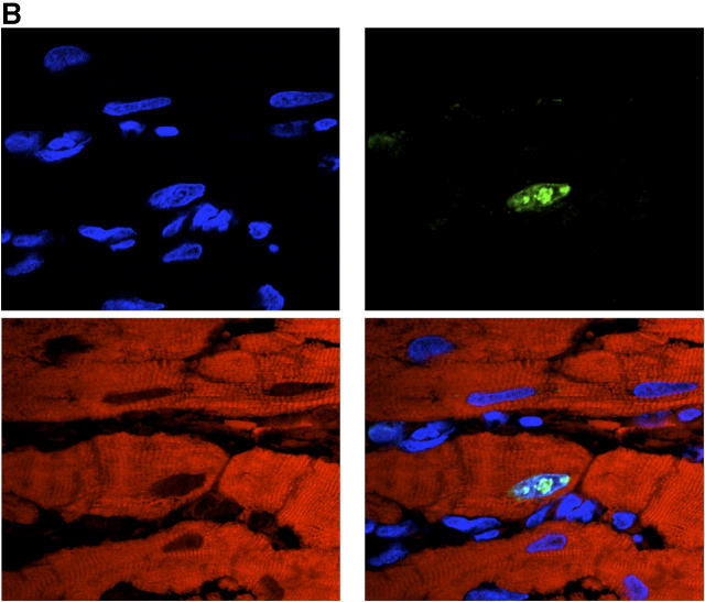
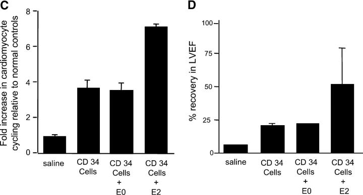
Effects of PAI-1 inhibition on regeneration of endogenous cardiomyocytes and on functional cardiac recovery. (A) Section from infarct of representative animal receiving CD34+ cells intravenously showing “finger” of cardiomyocytes of rat origin, as determined by expression of rat MHC class I molecules, extending from the peri-infarct region into the infarct zone is shown. These cellular islands contain a high frequency of myocytes staining positively for both cardiac-specific troponin I (brown) and rat-specific Ki-67 (dark blue; arrows). Sections from infarcts of representative animals receiving saline do not show same frequency of dual staining myocytes. (B) Confocal microscopy of peri-infarct tissue from representative animal receiving human CD34+ cells demonstrates cardiomyocytes whose nuclei (labeled blue by Cy5) concomitantly expressed Ki67 antigen (labeled green by FITC-conjugated secondary antibody reacting with polyclonal rabbit anti–rat primary antibody) and whose cytoplasm concomitantly stained for troponin I (labeled red by propidium iodide–conjugated anti-troponin mAb). (C) Animals receiving human CD34+ cells intravenously had a fourfold higher number of cycling cardiomyocytes at the peri-infarct region than that found in LAD-ligated controls receiving saline (P < 0.01). Combining intramyocardial E2 injection with intravenously delivered human CD34+ cells increased cardiomyocyte regeneration to levels 7.5-fold higher than in saline controls (P < 0.01). The scrambled DNA enzyme E0 had no such effect. (D) Combining intramyocardial injection of E2 with intravenous human CD34+ cells increased functional recovery of left ventricular ejection fraction at 2 wk from a mean of 22–50% (P < 0.01). The scrambled DNA enzyme E0 had no such effect. (C and D) Results are expressed as the mean ± SEM of three separate experiments.


