
Figure 2.
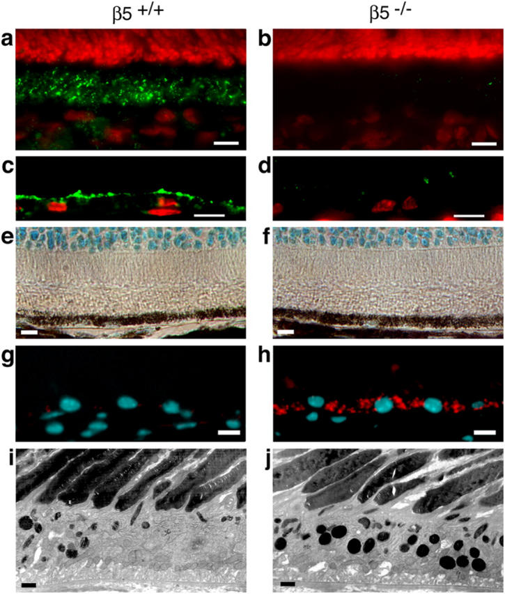
β5 integrin–deficient RPE accumulates autofluorescent lipofuscin with age. (a–d) Cryosections of 1-mo-old eyecups of wild-type (a and c) and β5−/− (b and d) mice were labeled with β5 antibody (green). Nuclei are shown in red. (a) In the wild-type mouse, whole retina sections with the neural retina present and β5 integrin was present in the photoreceptor outer segment layer, which also contains apical microvillar processes of RPE ensheathing photoreceptor outer segments (reference 28). (b) As expected, β5 integrin was absent from β5−/− retina. (c and d) To determine whether β5 integrin resided on photoreceptor outer segments or on RPE apical extensions, we prepared sections from eyecups from which the neural retina was peeled off before fixation. Prominent β5 integrin labeling at the apical surface of wild-type but not β5−/− RPE indicated that mouse RPE in vivo expresses apical αvβ5 integrin. (e–f) Methyl green–stained paraffin sections of eyecups from 12-mo-old wild-type (e) and β5−/− (f) mice were examined by light microscopy. Wild-type and β5−/− retina did not differ in appearance of photoreceptor nuclei (cyan at the top of the field), inner and outer segments (clear areas), and RPE (brown pigment and cyan nuclei). (g–h) Cryosections of eyecups from 12-mo-old wild-type (g) and β5−/− (h) mice were examined by wide-field fluorescence microscopy to detect DAPI-stained nuclei (cyan) and autofluorescence (detected in the rhodamine channel, red). β5−/−, but not wild-type, RPE contained numerous autofluorescent inclusion bodies resembling the characteristic age pigment lipofuscin in human RPE (32). (i–j) Ultrathin sections of eyecups from 12-mo-old wild-type (i) and β5−/− (j) mice were examined by transmission electron microscopy. Increased presence of electron dense inclusion bodies in β5−/− RPE compared with wild-type RPE confirmed the results obtained by fluorescence microscopy. Bars: (a–h) 10 μm; (i and j) 1 μm.