
Figure 3.
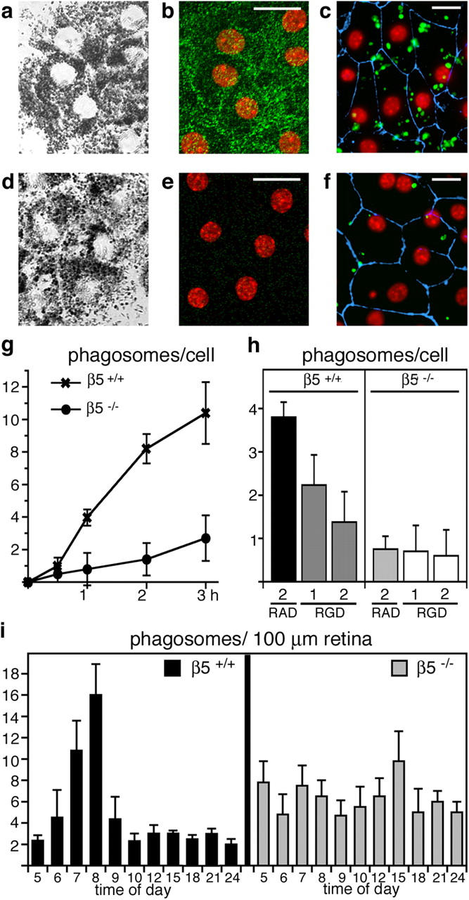
Lack of αvβ5 integrin impairs RPE phagocytosis of POS. We examined primary RPE in culture from wild-type (a–c) and β5−/− (d–f) mice. Confocal x-y scans of transmitted light (a and d) and of apical αvβ5 integrin (green) and nuclei (red) in the same fields (b and e) were used to compare general cell morphology and to illustrate apical αvβ5 receptors in wild-type, but not in β5−/−, RPE. (c and f) Junction marker ZO-1 (blue) and nuclei (red) appeared similar by wide-field fluorescence microscopy. However, β5−/− RPE in primary culture phagocytosed fewer FITC-POS (green) than wild-type RPE during a 1-h phagocytic challenge. (g) Quantification of in vitro phagocytosis assays showed reduced POS uptake by β5−/− RPE compared with wild-type RPE at all time points. Results represent means ± SD, n = 3, Student's t test, P < 0.01 at 1–3 h. (h) RGD peptides inhibited POS uptake by wild-type but not by β5−/− RPE in a concentration-dependent manner. Bars show 1-h FITC-POS uptake by wild-type and β5−/− RPE in the presence of 2 mM RAD-inactive control peptide, 1 or 2 mM integrin inhibiting RGD peptide as indicated. Since RGD peptides affected cell–substrate adhesion of both wild-type and β5−/− RPE at concentrations above 2 mM, these concentrations were not used for phagocytosis assays. Thus, the 63% inhibition we observed in the presence of 2 mM RGD peptide may not represent maximal possible inhibition of POS uptake by RGD peptides. Results represent means ± SD, n = 3, Student's t test, P < 0.01 for wild-type RPE, RGD versus RAD peptide at 2 mM. (i) Phagosome quantification in electron micrographs of retinal cross sections revealed that β5−/− retina lacked the characteristic burst of phagocytosis that followed the light onset (at 6.00 h) in wild-type retina. Mean (numbers of phagosomes) ± SD is shown. Sections from three eyecups per time point harvested in three separate experiments were examined.