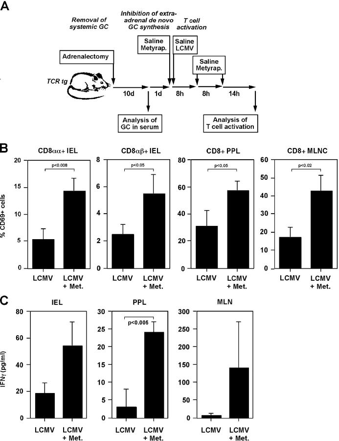
Figure 5.
In situ–produced GCs inhibit the activation of virus-specific intestinal T cells. (A) Schematic overview of the in vivo experiments. TCR tg mice were adrenalectomized to remove the major source of systemic GCs. After 10 d recovery, animals were treated with saline or metyrapone before infection with LCMV. Metyrapone or saline treatment was repeated after 8 and 16 h. 30 h postviral infection, the activation status of intestinal T cells were assessed. (B) Animals were treated as shown in A. IELs, PPLs, and mesentheric lymph node cells (MLNCs) were isolated. CD69 expression on the TCR tg T cells (Vα2+) on CD8αα and CD8αβ subsets (IEL), or CD69 expression on the TCR tg T cells on CD8+ T cells (PPL, MLNC) was monitored by flow cytometry. A typical experiment is shown (n = 3 per group; two experiments). Numbers indicate p-values of the Student's t test. (C) Animals were treated as described in A. IELs, PPLs, and MLNCs from LCMV-infected or LCMV-infected and metyrapone-treated (Met.) were isolated and cultured overnight. IFNγ in the culture supernatant was assessed by ELISA. Numbers indicate p-values.

