
Figure 2.
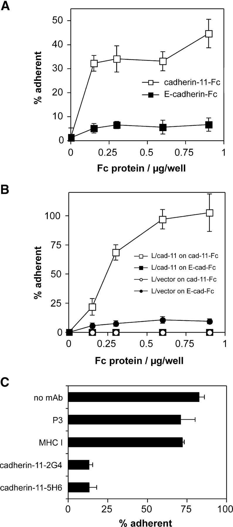
Cadherin-11–mediated adhesion of FLS and L cell transfectants. (A) Adhesion of cultured FLS derived from RA synovium to cadherin-11–Fc. Purified cadherin-11–Fc (open squares) or E-cadherin–Fc (closed squares) were immobilized on microtiter plate wells coated with polyclonal goat anti–human IgG antibody and blocked with BSA. The adhesion of RA-derived cultured FLS was determined in triplicate as described in Materials and Methods. (B) Specific adhesion of L cells to cadherin-11–Fc is conferred by cadherin-11 transfection. The assay was conducted as in A using control L cells (L/vector) or cadherin-11–transfected L cells (L/cad-11) and microtiter plates directly coated with the indicated concentrations of Fc proteins (cadherin-11–Fc or control E-cadherin–Fc). (C) Inhibition of adhesion of the L cell/cadherin-11 line to cadherin-11–Fc by anti–cadherin-11 antibodies. The assay was performed as in B but antibodies were preincubated with the cells for 10 min on ice. All the mAb were purified and used at 10 μg/ml and all mAb were mouse IgG1, except anti–mouse MHC-I (mouse IgG2a). The results are expressed as the mean percentage of cells that were adherent ±1 SD (n = 3).