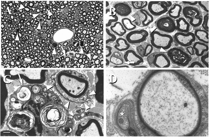
Figure 2.
Pathological features in sciatic nerves of GalNAcT−/− mice. (A) Toluidine blue-stained 1-μm epon section showing several myelinated fibers undergoing axonal degeneration (arrowheads) and a myelinated fiber surrounded by supernumerary Schwann cell processes (arrow). (Bar = 5 μm.) (B) Low-power EM image showing myelin figures and collapsed myelin at different stages of degeneration (arrowheads) and a thinly myelinated fiber surrounded by supernumerary Schwann cell processes (arrow) (Bar = 2.5 μm.) (C) EM image showing a macrophage containing myelin debris (arrow) and a minor onion bulb around a myelinated fiber (arrowheads point to Schwann cell processes). An endoneurial fibroblast also is apparent (∗) (Bar = 1 μm.) (D) EM image showing a thinly myelinated fiber and an axonal sprout (∗) invested by a Schwann cell (Bar = 200 nm). Mice were 12–16 weeks of age at the time nerves were harvested.

