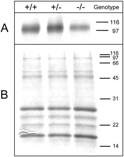
Figure 5.

Expression of MAG in GalNAcT−/− and control mice. Equivalent amounts of myelin from wild-type (+/+), GalNAcT+/−, and GalNAcT−/− mice (1.3 μg protein for immunoblotting or 5 μg protein for Coomassie staining) were subjected to SDS/PAGE (28). The positions of molecular mass standards are indicated in kDa. (A) Immunodetection of MAG. After resolution on a 10% polyacrylamide gel, proteins were transferred to a poly(vinylidene difluoride) membrane by using a semidry transfer apparatus. MAG, which migrates at 100 kDa, was detected by incubation of the blot with GenS3 mAb followed by an alkaline phosphatase-conjugated secondary antibody. Antibody binding was detected by development with nitro blue tetrazolium and 5-bromo-4-chloro-3-indolyl phosphate. (B) Staining of major myelin proteins. After resolution on an 8–16% polyacrylamide gradient gel, proteins were detected by using Coomassie brilliant blue stain. The major myelin proteins myelin basic protein (MBP) and proteolipid protein (PLP) are indicated. Mice were 15–23 weeks of age when brain tissue was collected for analysis.
