Figure 4.
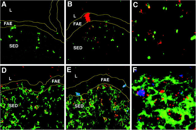
Reovirus infection in association with apoptosis in the PP SED. (A) PP cryosection from an uninfected control mouse stained with TUNEL (green) and examined by confocal microscopy. (B) PP cryosection from a mouse 24 h after peroral inoculation with T1L stained with TUNEL (green) and an antibody specific for reovirus σ1 (red). (C) Higher magnification of B showing reovirus σ1 in association with TUNEL+ inclusions. (D) PP cryosection from an uninfected mouse stained with antibodies specific for CD11c (green), activated caspase-3 (red), and reovirus σ1 (blue). Note that apoptotic material can be detected in both the FAE and SED. (E) PP cryosections from a T1L-infected mouse stained as in D. Note two reovirus-infected cells in the FAE that are also positive for activated caspase-3 as indicated by the purple color. (F) Higher magnification of the SED of E showing association of reovirus σ1 and activated caspase-3 as indicated by the purple color. L, lumen of small intestine; FAE, follicle-associated epithelium; SED, subepithelial dome. Borders of the FAE are indicated by yellow lines.

