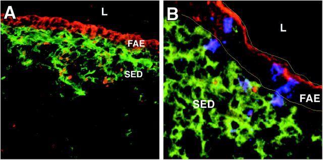
Figure 5.
Epithelial cell–derived cytokeratin in the FAE and SED of reovirus-infected mice. PP cryosections were prepared from mice 24 h after peroral inoculation with T1L. Sections were stained with antibodies specific for (A) CD11c (green) and cytokeratin (red), or (B) CD11c (green), cytokeratin (red), and reovirus σ1 (blue). Sections were examined by confocal microscopy. Cytokeratin can be detected in the DC region of the SED underlying the FAE. Reovirus σ1 shown in association with cytokeratin staining in the FAE and occasionally in the SED is indicated by the purple color. Borders of the FAE are indicated by yellow lines.

