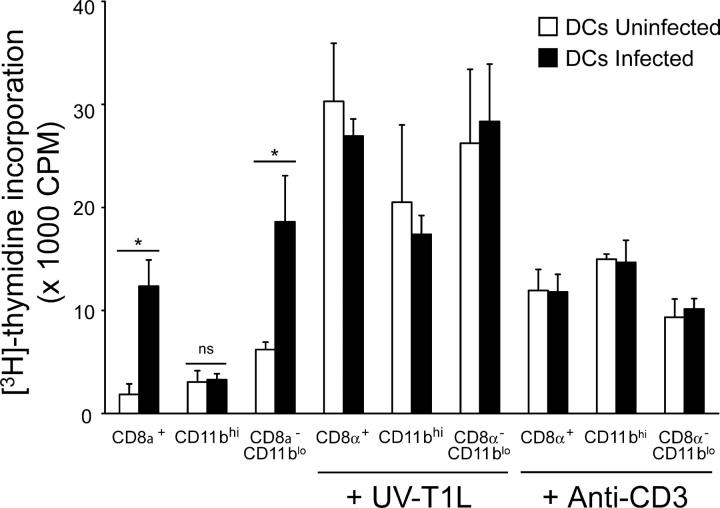
Figure 8.
Proliferation of T1L-primed CD4+ T cells induced by CD8α+, CD11bhi, and CD8α−/CD11blo subsets of DCs isolated from T1L-infected mice. DCs from uninfected (white bars) and infected (black bars) mice were purified by flow cytometry (>95% pure) and cocultured with T1L-primed T cells for 72 h at an APC/T cell ratio of 1:4 and in the absence or presence of UV-inactivated T1L (UV-T1L) or anti-CD3 as a positive control. Results are presented as mean [3H]thymidine incorporation. Error bars represent standard deviations for triplicate assays of one of two experiments performed with similar results. *, P < 0.05; ns, not significant.

