
Figure 5.
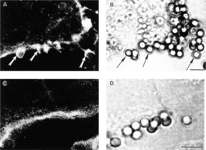
Confocal analysis of F-actin after IgG-coated particle binding to FcγR COS transfectants. Transfected COS cells were incubated with IgG-coated beads for 15 min at 37°C and were then fixed, permeabilized, and stained for F-actin using FITC-phalloidin. A and C show 0.5-μm–thick single confocal sections of F-actin staining, whereas B and D are companion nonconfocal transmitted light phase contrast sections. In A, an FcγRIIA transfectant shows F-actin extensions surrounding bound beads, whereas the parallel B provides the position of the beads. In C, an FcγRIA transfectant does not show an F-actin extensions or increases in F-actin due to target binding above background levels, whereas the parallel D shows the location of the beads. Cells were examined in all planes of focus to detect F-actin extensions as quantified in Table 4. Arrows, F-actin extensions in A, and associated beads in B. Bar, 5 μm.