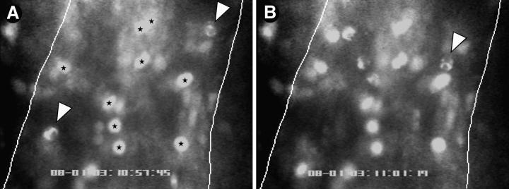
Figure 1.
Both PMNs and MNLs roll, but only MNLs stick in PLN-HEVs. Endogenous circulating WBCs were labeled in situ with the nuclear dye rhodamine 6G. Cells were recorded by fluorescence microscopy through an ×100 objective during their passage through HEVs. The micrographs show typical scenes in a segment of an order III venule (50-μm diameter, blood flow from top to bottom). Lines were drawn to demarcate lateral borders of the lumenal compartment. WBCs in the plane of focus show two distinct nuclear staining patterns allowing the identification of PMNs (irregular shape) and MNLs (homogenous round or oval staining pattern). (A) Two rolling PMNs (arrows) and several sticking MNLs (stars) can be seen. (B) 3.7 s later, one of the PMNs (A, lower left) has rolled out of the field of view, whereas the other PMN (arrowhead) has continued to roll slowly through the vascular segment. Sticking MNLs did not change their position. In the meantime, several additional rolling PMNs and MNLs entered the field of view.

