
Figure 1.
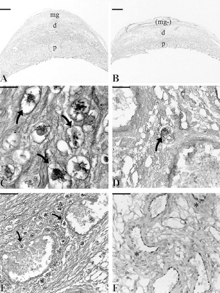
Comparison of day 12 placental morphology in NK cell– reconstituted tgε26 (A, C, and E) and tgε26 (B, D, and F) females. Placental (p) cross-sectional area measurements of NK cell–reconstituted tgε26 mice (NK+; A) were larger by 27–34% than gestational age-matched homozygous tgε26 mice (B). The NK+ females had well-developed metrial glands (mg) and higher cellularity in the decidua (d), whereas metrial gland development was absent (mg−) and the decidua was edematous in tgε26 females. When compared to control immune-competent females, NK+ females had 40–56% normal frequencies of uNK cells (arrows; C) whereas tgε26 females had 0–3% normal uNK cell frequencies (arrow; D). In the NK+ group of females, uNK cells (arrows) were frequently found surrounding decidual blood vessels and occasionally found within the vessel lumens (E). (F) demonstrates the decidual vessel anomalies found in tgε26 mice including thickened vessel walls (asterisks); A and B, bar = 1,000 μm; C and D, bar = 40 μm, E and F, bar = 100 μm; (A, B, and F) stained with hematoxylin and eosin; (C–E) stained with periodic acid– Schiff.