
Figure 4.
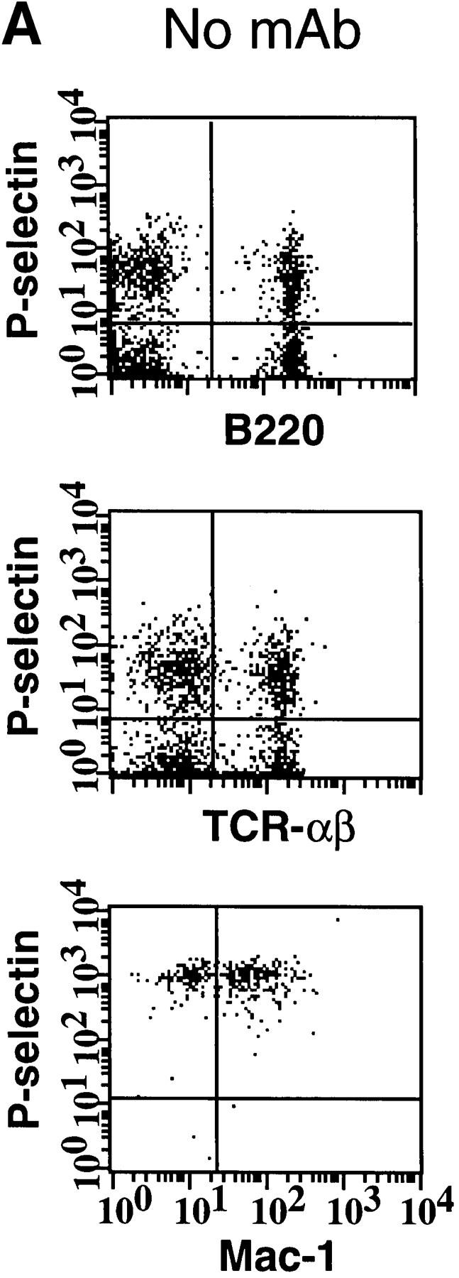
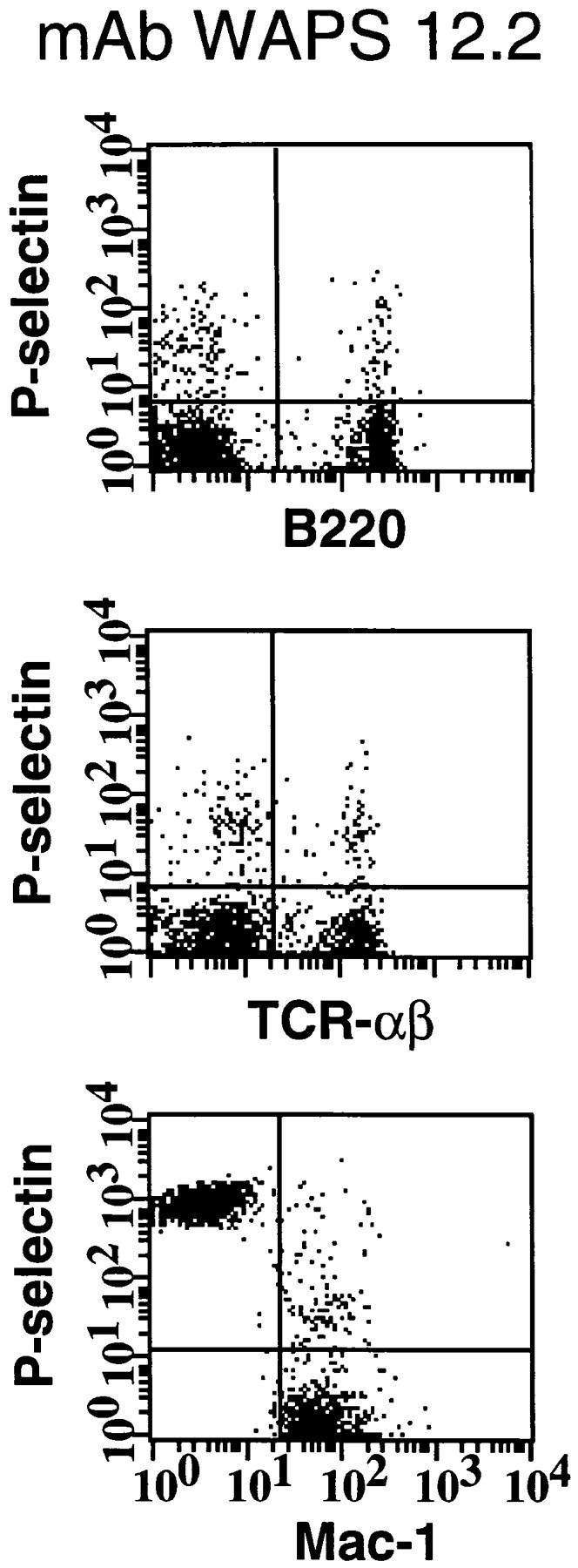

Activated human platelets bind to murine lymphocytes in suspension. Freshly isolated murine PBMCs were stained with FITC-conjugated antibodies to TCR-α/β, B220, and Mac-1 to identify T cells, B cells, and monocytes, respectively. Activated human platelets were stained with PE-conjugated nonblocking antibody to P-selectin and then mixed with labeled PBMCs. (A) Identification of PBMC subsets from L-selectin–deficient mice capable of aggregating with platelets as determined by two-color flow cytometry. The quadrant markers were drawn based on appropriate negative controls after gating separately for lymphocytes and monocytes. Double positive cells (upper right quadrant) represent the members of each subset capable of platelet binding. Events in the monocyte gate that show positive staining for P-selectin, but not Mac-1, are platelet aggregates (upper left quadrant). P-selectin–dependent adhesion was evaluated by treating platelets with mAb WAPS 12.2 before and during incubation with PBMCs. Similar results were obtained in wild-type mice. (B) Percentage of monocytes, T cells, and B cells of wild-type (+/+) and L-selectin-deficient (−/−) donor mice that interacted with activated human platelets. The effect of P-selectin blocking mAb WAPS 12.2 (open bar) and nonblocking mAb S12 (dotted bars) was evaluated as described in Materials and Methods. Bars reflect mean ± SD of four independent experiments.