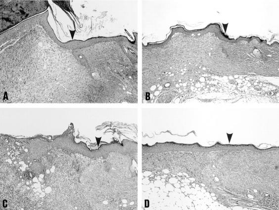
Figure 7.

Histological features of wound specimens from control C57BL/6 mice (A and B) and diabetic KK/Ta mice (C and D) 8 d after cutaneous wounding. NGF (1 μg; B and D) or vehicle solution alone (A and C) was applied to the wound space each day for 3 d beginning with the day of wounding as described in Materials and Methods. Sections were stained with hematoxylin and eosin. Original magnification: 60. Arrow heads, the original wound margin.
