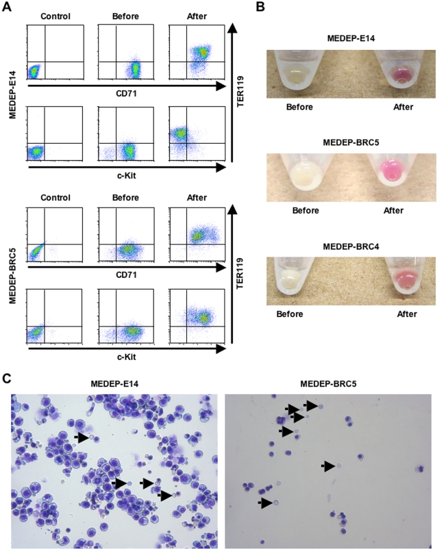
Figure 2. In vitro differentiation of MEDEP.
The in vitro differentiation of MEDEP-E14 was performed by culture for two days after deprivation of erythropoietin (EPO). The in vitro differentiation of MEDEP-BRC5 was performed by culture for three days after deprivation of stem cell factor (SCF) and addition of EPO. (A) Flow cytometric analyses. Control, results with isotype controls. Before and After, the cells before and after in vitro differentiation. CD71, transferrin receptor. c-Kit, receptor for SCF. TER119, a cell surface antigen specific for mature erythroid cells. (B) Cell pellets before and after in vitro differentiation. The method for in vitro differentiation of MEDEP-BRC4 is described in Figure S4. (C) Morphology of the cells after in vitro differentiation. Arrows indicate enucleated red blood cells. (A–C) Results shown are representative of three independent experiments.

