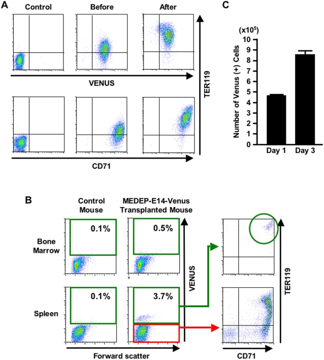
Figure 3. In vivo proliferation and differentiation of MEDEP.
A transformant of MEDEP-E14 expressing Venus as a marker was established, MEDEP-E14-Venus. (A) The in vitro differentiation of MEDEP-E14-Venus was performed by culture for two days after deprivation of erythropoietin. Control, results with isotype controls. Before and After, the cells before and after in vitro differentiation. (B) In vivo differentiation of MEDEP-E14-Venus cells. Acute anemia was induced in an immuno-deficient mouse (NOD-SCID) and the next day MEDEP-E14-Venus cells (2×10⁷ cells/mouse) were transplanted into the anemic mouse. Three days after cell transplantation, bone marrow and spleen cells were subjected to flow cytometric analyses. Control mouse, NOD-SCID mouse without cell transplantation. The vast majority of Venus-positive cells in the spleen show differentiation into CD71+TER119+ mature erythroid cells. (A, B) CD71 and TER119, see legend of Figure 2A. Results shown are representative of three independent experiments. (C) In vivo proliferation of MEDEP-E14-Venus cells. Cell transplantation was performed as in (B). We determined the proportion (%) of Venus-positive cells and calculated the absolute number of Venus-positive cells in the spleen. Day 1 and Day 3, one day and three days following cell transplantation, respectively. Values are mean±S.D. (N = 3).

